EUROPEAN FOULBROOD DISEASE TRANSMISSION AND REOCCURRENCE
Article originally published on the Canadian Honey Council Website, April 2023
https://honeycouncil.ca/wp-content/uploads/2023/04/Hivelights-Spring-2023.pdf
Identification of M. plutonius in hive matrices to understand European foulbrood disease transmission and reoccurrence.
Partly funded by the Canadian Bee Research Fund
Our team: Dr. Marta Guarna (PI; Agri-Food Canada), M.Sc. Heather Higo (President of the BCHPA), Dr. Sarah Wood (University of Saskatchewan) and Dr. Elemir Simko (University of Saskatchewan).
Report by
1. BC Technology Transfer Program, The British Columbia Honey Producers’ Association; nuriamorfin@ttp-bchpa.ca,
2. Department of Veterinary Pathology, Western College of Veterinary Medicine, University of Saskatchewan; midhun.jose@usask.ca, elemir.simko@usask.ca, sarah.wood@usask.ca, igor.moshynskyy@usask.ca
3. The British Columbia Honey Producers’ Association; heather.higo@gmail.com
4. Agriculture and Agri-Food Canada; marta.guarna@agr.gc.ca
European Foulbrood (EFB) is an infectious disease caused by the non-spore-forming bacterium Melissococcus plutonius, which infects 1–2-day old honey bee (Apis mellifera L.) larvae [1]. The infected larvae die rapidly after acquiring the infection by ingestion; the bacteria proliferate in the larvae’s gut [1]. The diseased larvae are characterized by a yellow or brown coloration, and they appear twisted within their cell [1]. Melisococcus plutonius can remain viable in brood cell walls and larvae faeces for years [2]. The transmission within the infected colony is through the fecal-oral route, mainly by worker bees feeding and cleaning diseased larvae [2]. Additionally, M. plutonius can be spread between colonies through robbing and drifting behaviours [3]. At a colony level, EFB is characterized by a non-uniform brood pattern and a foul smell when the infection is severe and a co-infection with secondary bacteria occurs [4].
The development of clinical signs of EFB has been linked to stress factors, including changes in nutrition; an association between EFB outbreaks and pollination of blueberries has been reported [5]. The treatment options for EFB are limited, although oxytetracycline is allowed Canada, its use is restricted to early spring and late fall to prevent honey contamination [6]. Additionally, the potential development of antibiotic resistant bacteria is an ongoing concern for international health authorities [7]. Control methods to prevent the spread of EFB include burning contaminated frames and hives, and using the ‘shook swarm’ method, in which bees are removed from contaminated frames and placed onto new equipment [8].
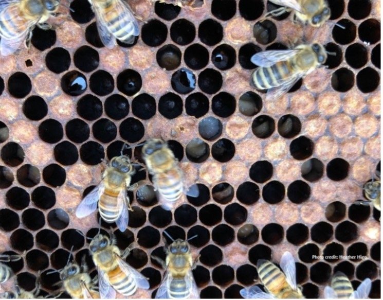

Figure 1a and b. Frames with larvae showing signs of EFB (Photo credit: Heather Higo and Sarah Wood).
EFB can remain as a covert infection, possibly due to mechanical contamination of honeycomb, facilitating subsequent outbreaks [4]. Additionally, beekeeping practices, such as the exchange of beekeeping equipment between operations has been pointed as a risky practice for the spread of the disease [4]. However, the identification of potential fomites (i.e. contaminated objects that can transfer infectious agents) responsible for the transmission of EFB within and between beekeeping operations is not well documented. The potential of spreading brood diseases through honeycomb is a growing concern, particularly in regions where EFB is endemic and causing increasing economic distress for beekeepers [9]. Therefore, preventing the spread of EFB between operations should be a high priority to control the disease. The identification of potential fomites is a first step towards prescribing advice on biosafety practices that could help prevent the spread of EFB between beekeeping operations, as has been done for other highly transmissible diseases in other types of animal production systems [10].

Figure 2. Colony morphology of M. plutonius. The bacteria were cultured in KSBHI agar
To assess the potential role of fomites in the transmission of EFB, 23 frames with nectar and pollen from honey bee colonies of two different beekeeping operations were collected in the Lower Mainland of British Columbia in the summer of 2022. Prior to the collection of frames, the colonies were used for blueberry pollination. Additionally, samples of larvae with visual signs of EFB were sampled (19 in total). M. plutonius was cultured from the swabs using KSBHI agar [11], and the cultured bacteria were later used for molecular identification using real time PCR (qPCR) [8]. Eighty four percent of the larvae with clinical signs of EFB were positive to both laboratory analyses: bacterial culture and qPCR. In addition, a technique to isolate M. plutonius from the hive matrices, including beeswax, nectar, and pollen is under development; successful identification of M. pluntonius with qPCR in samples of beeswax, honey, and pollen was achieved, and three positive cultures from beeswax were recorded. The confirmation of presence of M. plutonius in fomites will inform the design of future projects to study the transmissibility of M. plutonius, and later inform the development of biosafety practices to prevent the spread of EFB, such as targeting sanitation strategies to fomites with the highest risk of disease transmission.
Acknowledgement: This research was partly funded by The Canadian Bee Research Fund to MMG – AAFC J-002912. We gratefully acknowledge the financial support of the B.C. Ministry of Agriculture and Food to the BC Technology Transfer Program. We also thank the participating beekeepers for their valuable contribution.
References
Forsgren, E. (2010). European foulbrood in honey bees. Journal of Invertebrate Pathology, 103, S5-S9.
Forsgren, E. et al. (2013). Standard methods for European foulbrood research. Journal of Apicultural Research, 52(1), 1-14.
McKee, B.A. et al. (2004). The transmission of European foulbrood (Melissococcus plutonius) to artificially reared honey bee larvae (Apis mellifera). Journal of Apicultural Research, 43(3), 93-100.
Vidal-Naquet, N. (2015). Honeybee Veterinary Medicine: Apis mellifera L. 5M Publishing.
Olmstead, S. et al. (2019). Evaluating the effect of feeding pollen substitute to honey bee colonies destined for wild blueberry pollination in Colchester County, Nova Scotia. Nova Scotia. Available from https://www.perennia.ca/wp-content/uploads/2019/10/ATTTA-FactSheet-Oct-2019.pdf [accessed on 17 February 2020].
Pernal, S.F. & Clay, H. (2013). Honey bee diseases and pests, 3rd Edition. Canadian Association of Professional Apiculturists, Beaverlodge, AB, Canada 68 pp.
Chang, Q. et al. (2015). Antibiotics in agriculture and the risk to human health: how worried should we be?. Evolutionary Applications, 8(3), 240-247.
Budge, G.E. et al. (2010). The occurrence of Melissococcus plutonius in healthy colonies of Apis mellifera and the efficacy of European foulbrood control measures. Journal of Invertebrate Pathology, 105(2), 164-170.
Gregoris, A. et al. (2020) European foulbrood may affect availability of honey bee colonies in pollination-deficient blueberry farms. Entomology 2020, 68th Annual Meeting of the Entomological Society of America, virtual meeting. 11-25 November 2020.
Karus, A. et al. (2018). Biosafety and biosecurity manual. EMU DSpace. Available from https://dspace.emu.ee/xmlui/handle/10492/5453?locale-attribute=en [accessed on November 20, 2021].
Arai, R. et al.. (2012). Diversity of Melissococcus plutonius from honeybee larvae in Japan and experimental reproduction of European foulbrood with cultured atypical isolates. PloS one, 7(3), e33708.